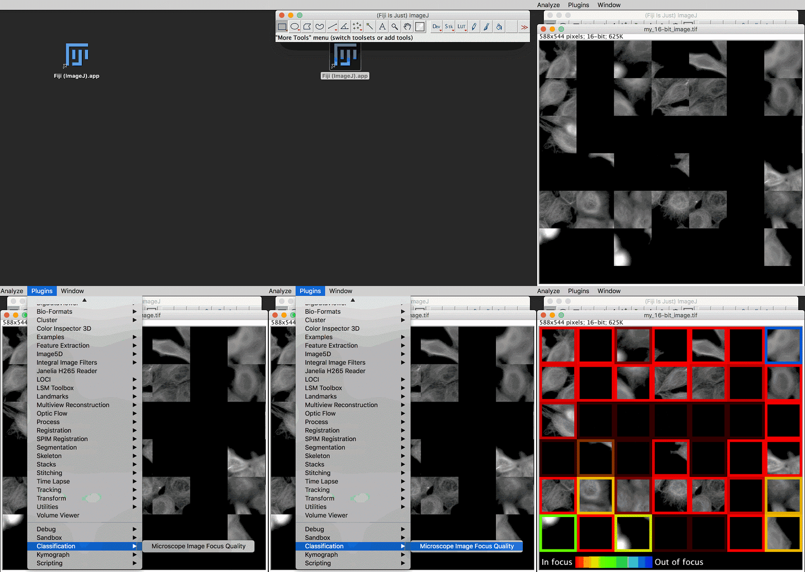
Editor's Note: Although modern microscopes are equipped with advanced hardware systems, the large image datasets they generate often contain low-quality or out-of-focus images. To ensure accurate and unbiased scientific analysis, it is essential to use high-precision automated image processing techniques. Google researchers have developed deep learning models to address this challenge, enabling scientists to capture and analyze high-quality images more efficiently. Below is a refined version of the original content.
Many scientific imaging applications, particularly in microscopy, produce terabytes of data daily. These datasets benefit greatly from recent advances in computer vision and deep learning. As we collaborate with biologists on robotic microscopy projects, we’ve discovered that cleaning up noisy or out-of-focus images is both challenging and crucial for reliable data. Moreover, we've realized that many scientists may not be fluent in coding, yet they still want to leverage deep learning for image analysis. One specific issue we aim to solve is handling out-of-focus images. Even with state-of-the-art autofocus systems, misconfiguration or hardware incompatibility can lead to poor image quality. Automated focus evaluation helps detect, troubleshoot, and remove such problematic images.
Secured from Deep Learning
In the article "Assessing Microscope Image Quality with Deep Learning," we trained a neural network to evaluate the focus quality of microscope images more effectively than previous methods. We also integrated a pre-trained TensorFlow model into Fiji (ImageJ) and CellProfiler—two widely used open-source tools for scientific image analysis. These tools can be accessed via a graphical interface or through scripting, making them accessible to a broader range of users.

The workflow of our machine learning project is detailed in published papers and open-source code (including TensorFlow, Fiji, and CellProfiler). We assembled a training dataset by defocusing 384 cell images, eliminating the need for manual labeling. We then trained a model using data augmentation to improve generalization, including an unseen cell type captured from a different microscope. Finally, we deployed a pre-trained model, which doesn’t require user-defined parameters and offers more accurate focus assessments. For better interpretability, the model evaluates focus quality on 84×84 pixel blocks, represented as colored borders in the image above.
What About Images Without Target Objects?
One interesting challenge we faced was dealing with image patches that were "blank" — meaning they contained no target objects. In such cases, there’s no meaningful concept of focus quality. Instead of manually labeling these blank patches, we allowed the model to identify them. However, we configured the model to predict a probability distribution of defocus levels, enabling it to express uncertainty in those regions.
What’s Next?
Deep learning-based approaches for scientific image analysis are set to improve accuracy, reduce manual effort, and potentially lead to new discoveries. The value of shared datasets, effective models, and user-friendly tools highlights the importance of widespread adoption across all scientific fields. As these technologies continue to evolve, their integration into everyday research practices will become increasingly vital.
The disposable ecig arms race continues, with ever larger devices continuing to hit the market. The newest disposable juggernaut is the IGET XXL, with a nic strength of 5 percent, this device holds over five times more nic salts than a standard disposable bar and approximately nine times more than a Juul Pod. What this means is written boldly on the packaging for all to see: 1800 puffs, a number that will vary depending on how long your average inhale is.
IGET,Disposable E-Cigarette ,Disposable Light Vape,Disposable Device Pen,Vape Pod Smoke
Shenzhen Ousida Technology Co., Ltd , https://en.osdvape.com
