
Author: Murata Electronics Ulf Meriheinä
Through the design of acceleration sensors and gyroscopes based on microelectromechanical system (MEMS), MEMS technology has been widely used in the field of navigation and game software; however, micro electromagnetic sensor technology is increasingly used Used in the medical field.
MEMS is commonly used in patient diagnostic equipment. This diagnostic instrument is used to detect the function of the patient's heart. Medical staff usually use electrocardiogram to check the patient's heart function. During the electrocardiogram examination, the medical staff will connect a set of electrodes to the human body to make it contact with the skin surface. In this way, we can measure complex vector electrocardiogram (VCG). Vector ECG is a traditional method, which can record the amplitude and time of the P-QRS-T wave of the patient's ECG or only the time of the peak of the R wave. This vector electrocardiogram is the same as the image displayed in the heart rate monitor or exercise computer shown in Figure 1.
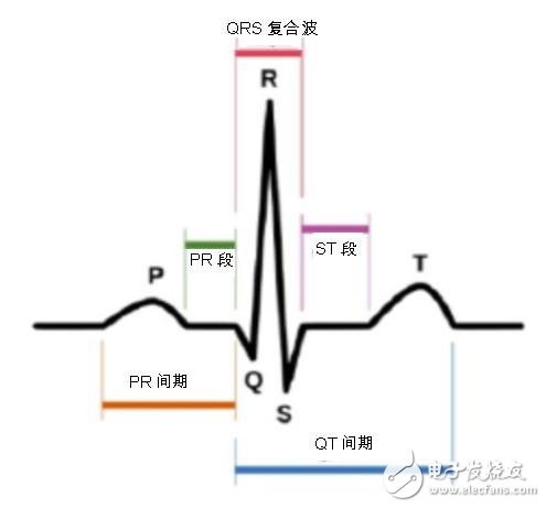
Figure 1: P-QRS-T wave in electrocardiogram
The electrocardiogram can provide us with a lot of relevant information, including cardiac dysfunction, heart disease, and recovery of cardiac function, the patient's physical and psychological stress. However, the electrocardiogram cannot well check the mechanical blood pumping function of the patient's heart or the function of the heart. In addition, these electrodes may interfere with the patient's daily life, especially interfere with the patient's recreational activities and night ECG monitoring. Fortunately, in the medical field, we can check the function of the heart by other means, such as cardiac ultrasound monitoring and cardiac shock scan (Ballistocardiology, BCG). Mechanical cardiac shock scans record the electrical signal of the heart, but its signal is delayed by 30 to 40 microseconds.
Using the heart shock scan, we can record the mechanical activity of the heart by detecting the power and acceleration of the chest. In addition, there is a method to detect the pumping function of the heart by remotely controlling the heart shock scan. . Using remote-controlled cardiac shock scans, we can eliminate the need for electrodes or instruments to touch the patient's skin surface. This method is a great advantage for detecting patients' ECG activities in daily life. Since blood flow usually flows in one direction, we can only record the blood flow direction on one axis, such as the length of a person from head to toe.
Cardiac shock scans can be well used in the field of preventive medicine, such as to detect physical and psychological stress conditions or to detect early coronary heart disease. The amplitude shown in Figure 2 measures the stroke volume of the heart: from a time point of view, we can infer the function of the heart, heart rate, and heart rate variability. The variability of the heart rate is a good record of the patient's recovery and the pressure experienced during the test. The amplitudes I and IJ in Figure 2 can be effectively used to assess the severity of certain diseases, such as valvular heart disease, coronary heart disease, and even the life span of patients.
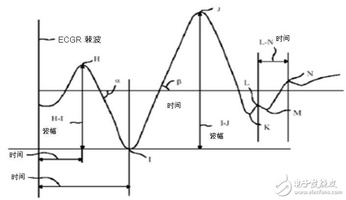
Figure 2: Waveform of cardiac shock scan.
It is very difficult to record the shock signal of the heart through the acceleration sensor. The acceleration signal of the heart activity is very weak, and the sensor itself has relatively high noise and environmental noise. At the same time, there may also be mechanical frequency response and vibration during the medical examination Interference (such as electronic scales for measuring weight and patient beds). It is especially important to monitor cardiac shock scans in hospital beds, because such cardiac shock scans can monitor the patient's physical health and functional recovery without disturbing the patient's sleep. The cardiac shock scan does not require electrode contact, so it does not affect the patient's daily activities and does not interfere with the patient's sleep. Based on this unique advantage, cardiac shock scans play a significant role in certain areas, including monitoring the heart function of patients suffering from sleep disorders due to physical or mental illness, and monitoring athletes ’best training results to avoid Overtraining.
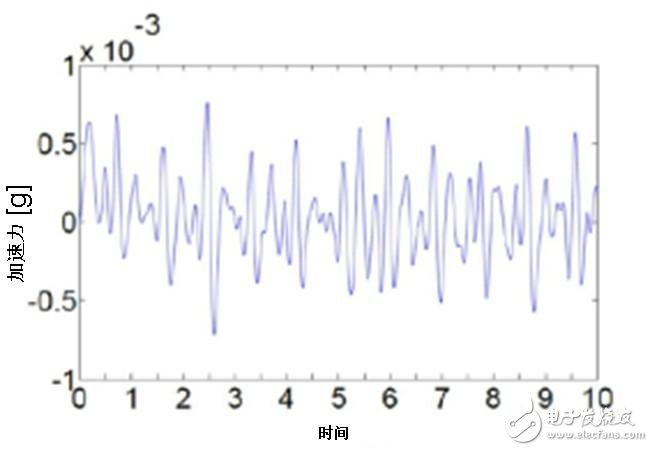
Figure 3: The accelerometer SCA121T from Murata, Japan, measures the waveform of the patient's cardiac shock scan from the hospital bed.
Diameter 30mm SPI 3D Tube , it also called 3d light and 3D Led Light Tube, we have different length 0.5m 3D tube, 1m 3D tube, 1.5m 3D tube and 2m 3D tube, the SPI 3D Led Tube can use for led dancing light. SPI LED Tube Light is similar DMX 3D LED Tube , but the control ic is different.
Diameter 20mm SPI 3D Tube, it also called meteor lights and Led Video Light , we have different length 0.5meters 3D led tube, 1meter 3D led tube, 1.5meters 3D led tube and 2meters 3D led tube , the SPI 3D LED Tube can use for led dancing light. Spi Led Tube Light is similar DMX 3D LED Tube, but the control ic is different.
Photo show of SPI 3D Tube:


Spi 3D Led Tube Light,3D Led Dancing Light,3D Led Light Tube,Led Video Light
Shenzhen Iseeled Technology Co., Ltd. , https://www.iseeledlight.com